A note to potential referring doctors
Complete decongestive therapy (CDT) and vestibular rehabilitation are in no way meant to supercede the care of a medical doctor.
Medical evaluation is needed, of course, to rule out other possible causes for the patient’s complaints, to confirm the presenting diagnosis, and to manage the patient’s overall health. Vestibular rehabilitation and CDT are services typically delegated by doctors to appropriately trained and experienced ancillary professionals including but not limited to physio- and occupational therapists, registered nurses, and audiologists.
CDT for lymphedema specifically is a labor-intensive and supply-heavy treatment requiring multiple, at times logistically complicated, visits per week. This is not compatible with most doctors’ busy schedules and preferred working methods. I am able to provide these specialized treatment services for your patients.
Upon evaluation at CLTVR, I will send a full report to the prescribing doctor’s office as well as progress reports as needed over the course of treatment. Your patient’s progress will be tracked with objective, validated tests and measures. After the patient’s specific complaints are resolved to the best of my ability, the patient will be advised to continue for regular medical follow up as needed with the prescribing doctor. Physiotherapy-based treatment of edema, cancer-related functional limitations, dizziness, and imbalance are the only services provided at CLTVR. We are here to support you in managing these specific symptoms for your patients who have these challenging diagnoses.
Nathan Bridgeman, PT, CLT-LANA
Physiotherapist, Certified Lymphedema Therapist- Lymphology Association of North America
A note on compression garments
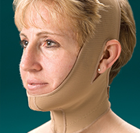
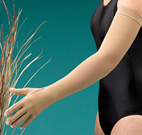
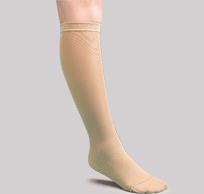
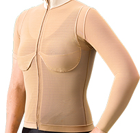
| Every patient with lymphedema needs to be provided with an appropriately fitted compression garment. This is not a controversial statement. This is the consensus recommendation of lymphology experts worldwide, as is clearly demonstrated in the literature linked below. I have heard the objection raised that people in Hong Kong, due to the subtropical climate and cultural norms of vanity, will never agree to this course of care. Based upon my past practice and correspondence with international lymphedema specialists, I can offer assurances that no one, anywhere, wants to wear a compression garment. The prospect of relying upon compression garments every day for the rest of one’s life is not a pleasant one. Consider, however, that newly diagnosed diabetics do not want to inject insulin; people with heart failure associated with sleep apnea do not want to sleep wearing a CPAP mask; drug addicts do not want to stop using drugs; women with breast cancer do not want to have mastectomies -- the world of medicine is filled with such examples of undesirable, but necessary, treatments. It is the duty of a practitioner treating a patient with lymphedema to impress upon the patient the reality of the situation, not to provide them with a more appealing, but insufficient, alternative. Imagine if doctors and surgeons did not press upon patients the need for insulin or surgery simply because they are unpleasant. I implore any doctor reading this to ensure that every suspected lymphedema patient you encounter be referred to a qualified lymphedema therapist, whether at CLTVR or elsewhere. Nathan Bridgeman, PT, CLT-LANA |
  |
||
 |
.jpg) |
 |
 |
Reviews of the Evidence
Diagnoses treated with complete decongestive therapy, and modifications of the technique
Lymphedema and Complete Decongestive Therapy
Further Information on the Physiology of the Lymphatic System: Its Function and Dysfunction
Diagnoses treated with balance therapy, vestibular rehabilitation, and related techniques
Vestibular Rehabilitation
| Reviews of the Evidence | |
|---|---|
| I recognize that the highly specific treatment regimens which I provide for relatively uncommon and non-life-threatening conditions are not among those to which many doctors are exposed in medical school, or even over a long and varied practice. Below please find a brief list of links to some of the more comprehensive peer-reviewed systematic literature reviews and specialist-organization position papers supporting the use of CDT and vestibular rehabilitation as the methods of choice for treatment of their respective diagnoses and symptom sets. I have a number of pre- and post-treatment photographs showing the effects of CDT in actual patient cases. I do not believe it is appropriate to post such images on a publicly-accessible website but I will provide copies to doctors upon request. CDT Compression Garments CDT Training of Certified Lymphedema Therapists Compression Garments Vestibular Rehabilitation for Vestibular Hypofunction Canalith Repositioning Maneuvers for BPPV Canalith Repositioning Maneuvers for BPPV |
|
| Diagnoses treated with complete decongestive therapy, and modifications of the technique: | |
| Patients with damage to axillary lymph nodes: Breast Cancer, Upper Body Melanoma Patients with damage to inguinal or abdominal lymph nodes: Gynecological and Urological Cancers, Rectal Cancer, Lower Body Melanoma Patients with damage to cervical lymph nodes: Pharyngeal Cancer, Head and Neck Melanoma Secondary Lymphedema of Non-Oncological Etiology Primary Lymphedema Lipedema (Total limb volume is not as amenable to reduction as in the case of lymphedema, but symptoms of discomfort improve with CDT and fitting with compression garment) Venous Disease (Typically easier to treat and faster to reduce than lymphedema. Compression garments remain the standard of care for long term management) |
|
| Lymphedema and Complete Decongestive Therapy | |
| Lymphedema Anyone who has had surgery or radiation to the lymph nodes in their axilla, groin, abdomen, or neck is at risk of developing lymphedema7,8, sometimes years after cancer treatment has concluded8. Various studies have found lymphedema affecting between 12%-60% of breast cancer survivors8-11. Even lymph node sparing sentinel node biopsy was found to carry a 4%-6% risk of developing lymphedema,12 with one study finding a 17% lymphedema rate in this population13. Lymphedema has been found to affect the legs of 28%-47% of survivors of gynecological cancers8,14,15. Between 12% and 75% of patients treated for head and neck cancer16-20 have been found to suffer with a particularly disfiguring and challenging to treat21 form of lymphedema in this part of the body. The wide range of findings in the above studies speak to the fact that lymphedema remains a condition relatively neglected by contemporary medicine: they result largely from researchers disagreeing on standardized criteria of diagnostic definition and methods and timing of measurement7. |
|
| Complete Decongestive Therapy (CDT)7,8 Complete decongestive therapy (CDT) is the internationally recognized standard of care for lymphedema. It is not one alternative among many. Lymphedema is a condition whose management requires specialized training and experience above and beyond basic schooling in physiotherapy or nursing. Specialized training allows the practitioner to best assess the patient’s condition and response to treatment as well as to identify complications in the progression of the disease. There are two phases of CDT treatment. Phase one is the active treatment phase, done in the clinic. In phase one, edema is rapidly reduced by coordinated treatment. Phase one ends when the patient is fit with an appropriate compression garment and he or she is able to perform all self-care activities. Phase two is the maintenance phase: lifestyle changes the patient must make to control their disease, analogous to a diabetic needing to change his or her diet and inject insulin. In short, CDT has four components: Because lymphedema results from a “low-output” failure of the lymphatic system, edema formation is a continuous process: as long as blood is flowing, fluid is being released into tissues and accumulating due to the body’s inability to remove it. This requires a continuous counteracting force. The wearing of compression garments throughout daytime activity provides this counteracting force. There is no substitute for this vital element of treatment. Coordination of care is important to best control edema. If a patient can not be seen for several days such that their compression bandages are removed between treatment sessions, the edema re-accumulates and his or her lymphedema will be poorly controlled. If there is a delay between measuring for the compression garment and obtaining it, compression bandages must be maintained until the compression garment is obtained, or the edema can re-accumulate to the point that the measured garment no longer fits appropriately when it arrives. Once edema is reduced in phase one, compression must be maintained or the edema will return. A patient can easily relapse to their starting condition solely due to their failure to comply with the use of compression garments. A course of intensive treatment for lymphedema is wasted time, wasted effort, and wasted healthcare resources if it does not end with the patient’s use of a properly fit compression garment. |
|
|
1.Tobin MB, Lacey HJ, Meyer L, Mortimer PS. The psychological morbidity of breast cancer-related arm swelling. Cancer 1993; 72(11): 3248-52. 2.De Godoy JMP, Braile DM, de Fatima Godoy M, Longo O Jr. Quality of life and peripheral lymphedema. Lymphology 2002; 35(2): 44- 50. 3.Williams AF, Moffatt CJ, Franks PJ. A phenomenological study of the lived experiences of people with lymphoedema. Int J Palliat Nurs 2004; 10(6): 279-86. 4.McWayne J, Heiney SP. Psychologic and social sequelae of secondary lymphedema: a review. Cancer 2005; 104(3): 457-66 5.Hayes SC, Janda M, Cornish B, Battistutta D, Newman B. Lymphedema after breast cancer: incidence, risk factors, and effect on upper body function. J Clin Oncol 2008; 26: 3536-42. 6.Mortimer PS. Managing lymphedema. Clin Exp Dermatol 1995; 20(2): 98-106. 7.Foldi M, Foldi E. Lymphostatic diseases. In: Strossenruther RH, Kubic S, editors. Foldi’s textbook of lymphology for physicians and lymphedema therapists. 2nd edition. Munich, Germany: Urban and Fischer; 2006. pp. 224–240. 8.Lymphoedema Framework. Best Practice for the Management of Lymphoedema. International consensus. London: MEP Ltd, 2006. 9.Schrenk P, Reiger R, Shamiyeh A, Wayand W. Morbidity following sentinel lymph node biopsy versus axillary lymph node dissection fro patients with breast carcinoma. Cancer 2000; 88(3): 608-14. 10.Meric F, Buchholz TA, Mirza NQ, et al. Long-term complications associated with breast-conservation surgery and radiotherapy. Ann Surg Oncol 2002; 9(6): 543-49. 11.Ozaslan C, Kuru B. Lymphoedema after treatment of breast cancer. Am J Surg 2004; 187(1): 69-72. 12.Golshan M, Martin WJ, Dowlatshahi K. Sentinel lymph node biopsy lowers the rate of lymphedema when compared with standard axillary lymph node dissection. Am Surg 2003; 69: 209-11. 13.Francis WP, Abghari P, Du W, Rymal C, Suna M, Kosir MA. Improving surgical outcomes: standardizing the reporting of incidence and severity of acute lymphedema after sentinel lymph node biopsy and axillary lymph node dissection. Am J Surg 2006; 192: 636-9 14.Hong JH, Tsai CS, Lai CH, et al. Postoperative low pelvic irradiation for stage I-IIA cervical cancer patients with risk factors other than pelvic lymph node metastasis. Int J Radiat Oncol Biol Phys 2002; 53(5): 1284-90.nomenological study of the lived experiences of people with lymphoedema. Int J Palliat Nurs 2004; 10(6): 279-86. 15.Ryan M, Stainton MC, Slaytor EK, et al. Aetiology and prevalence of lower limb lymphoedema following treatment for gynaecological cancer. Aust N Z J Obstet Gynaecol 2003; 43(2): 148-51. 16.Buntzel J, Glatzel M, Mucke R, Micke O, Bruns F. Influence of amifostine on late radiation-toxicity in HNC-a follow-up study. Anticancer Res 2007; 27:1953–1956 17.Dietz A, Rudat V, Nollert J, Helbig M, Vanselow B, Weidauer H. Chronic laryngeal edema as a late reaction to radio-chemotherapy. HNO 1998; 46:731–738 18.Schiefke F, Akdemir M, Weber A, Akdemir D, Singer S, Frerich B. Function, postoperative morbidity, and quality of life after cervical sentinel node biopsy and after selective neck dissection. Head Neck 2009; 31:503–512 19.Wolff HA, Overbeck T, Roedel RM et al. Toxicity of daily low dose cisplatin in radiochemotherapy for locally advanced HNC. J Cancer Res Clin 2009; 135:961–967 20.Deng J, Ridner S et al. Prevalence of Secondary Lymphedema in Patients With Head and Neck Cancer. J. Pain Symptom Manage 2012; 43(2):244-252 21.Smith B, Lewin J Lymphedema management in head and neck cancer. Curr Opin Otolaryngol Head Neck Surg 2010; 18:153–158 |
|
| Further Information on the Physiology of the Lymphatic System: Its Function and Dysfunction | |
| The lymphatic system is a part of the circulatory system with an important function in maintaining the health of body tissues, regulating the fluids and other substances in our bodies outside of the blood, and in fighting infection and disease. It is a network of vessels responsible for transporting fluid and substances too large to travel in the blood out of distant parts of the body, cleansing and filtering this fluid, and returning it back to the blood supply. The lymphatic system can be thought of as a “housecleaning” or “sewage” system for the microscopic environment between the body’s cells. When the lymphatic system is damaged, this can manifest in a variety of problems commensurate with the system’s many functions. Nutrients, oxygen, and other vital substances must leave the blood capillaries and enter the interstitial space between the body’s cells dissolved in fluid called “ultrafiltrate”. The ultrafiltrate washes over the cells which take what substances they need and excrete those they need to rid themselves of. The bulk of the ultrafiltrate, about 90%, is reabsorbed into the capillary as the blood prepares to enter the venous system and begin its journey back to the heart. About 10% of the ultrafiltrate fluid, including substances too large to travel in the blood capillaries (but still microscopically small) such as certain proteins, dead body cells, debris, or bacteria must return to the heart by means of the lymphatic system. This 90/10 ratio is fairly constant. The more blood passing through a capillary, the more ultrafiltrate remains for the lymphatic system to transport away. The forces at play here are important. There is, at the arterial end of the capillary (end of the capillary closer to the artery), a great deal of mechanical pressure from the pumping action of the heart. This tends to squeeze the ultrafiltrate out of the capillary and into the interstitium. At the venous end of the capillary (end of the capillary closer to the vein), those substances, such as proteins and electrolytes, which have remained behind in the blood tend to exert a negative osmotic pressure. This tends to pull the fluid back into the capillary. Unlike the circulation of the blood, lymphatic vessels provide their own pumping action rather than being pumped by the heart. Each lymphatic vessel is divided lengthwise into innumerable tiny segments separated by one-way valves and wrapped around with smooth muscle cells which rhythmically squeeze to push the fluid, now called lymph, through the valve and on into the next segment of the vessel. In order to accommodate increased accumulation of ultrafiltrate, such as in the case of increased blood flow to the area, the frequency of this rhythmic pumping can increase up to ten times the resting rate. As the lymph travels towards the center of the body, it is routed into lymph nodes which exist throughout the body but are clustered in groups at the major joints, the neck, and in the gut. The body is “geographically” separated into areas whose lymph drains into one of these clusters: the arm and half of the chest and back drain into the armpit; the leg and half of the lower body drain into the groin; parts of the lower abdomen and inner thighs drain into the gut; and the head and neck drain into the neck. Because only a specific set of nodes is damaged in cancer treatment, only a specific body region becomes at risk of developing lymphedema. A breast cancer survivor is not at risk of developing lymphedema in her opposite side arm, neck, or legs. A nasopharyngeal cancer survivor is not at risk of developing arm or leg lymphedema. Not everyone who has had damage done to their lymphatic system proceeds to develop lymphedema. The physiological processes involved in the development of lymphedema are not fully understood. It is theorized that the vessels of a damaged lymphatic system begin to pump at a higher resting speed, thus allowing a smaller number of functioning nodes and vessels to do the job of a healthy lymphatic system. This can only work up to a point: there is a limit to the rate at which the vessels can pump, beyond which fluid will begin to accumulate in the interstitium. Most of the common-sense lymphedema risk-reduction strategies are based on the concept of minimizing increases in local blood circulation and therefore ultrafiltrate production. As lymphedema progresses, several changes occur in the body. Overloaded lymphatic vessels begin to stretch out as they are filled beyond capacity with fluid. This stretching pulls the valves apart, in time irreversibly damaging the vessel’s ability to function. Ultrafiltrate, still being formed as a natural part of the circulatory process, accumulates in the interstitium. The ultrafiltrate, recall, contains dissolved substances such as proteins and electrolytes. These substances tend to exert a negative osmotic pressure in the interstitium. This change of the relative pressures inside and outside of the blood capillary upsets the natural balance of forces and causes more ultrafiltrate to leave the blood and less ultrafiltrate to reabsorb into the blood, which, in turn, puts a greater strain on the lymphatic system. These sorts of “feed-forward” mechanisms help explain why lymphedema tends to get worse over time. Tissues swell as the fluid accumulates without an outlet. Cells are physically pushed further away from the capillaries which supply their nutrients and remove their waste. Some of the proteins in the ultrafiltrate are, on a microscopic scale, like long sticky threads. As these proteins accumulate they begin to stick together randomly, similarly to the way wool will mat into felt. Also in the interstitium are cellular waste products and other chemicals, such as inflammatory mediators which, under normal conditions, are removed by the lymphatic system but, instead, are being left to accumulate. These processes, along with chemically-mediated fibroblast proliferation, cause fibrosis of the soft tissues. Fibrosis first causes the soft tissues of the body to develop a spongy, wet clay-like texture which will “pit” in response to mechanical pressure. Over time the proteins can become so densely packed that normally soft body tissues can become hard to the touch. Fibrotic tissue, chronic inflammation, and the increased distance between the blood supply and the body’s cells all work together to make the interstitium a less healthy environment for cells to live. Nutrients have difficulty diffusing through the crowded interstitium to reach cells. Many bacteria, on the other hand, find the environment to be very conducive to life: with protein and weakened cells to serve as food. The usual defense mechanism, of immune cells in the lymph nodes being alerted to the presence and nature of an infection and beginning the production of antibodies, is impaired. This part of the body is at increased risk of infection. All of the above has important implications for the treatment of lymphedema. The mechanical pressure inside the capillary pushes fluid out into the interstitium. Osmotic pressure inside the capillary pulls fluid back into the capillary. In lymphedema, an abnormal osmotic pressure in the interstitium pulls more fluid into the interstitium. The use of a compression garment adds a new force into this equation. An external mechanical pressure is applied which manifests upon the interstitium causing an increased mechanical pressure in the interstitium which tends to push fluid back into the capillary. Less ultrafiltrate is formed and more ultrafiltrate is reabsorbed for that time that the garment is worn. This explains one important reason why the use of alternate treatments is not an adequate substitute for the use of compression garments. Ultrafiltrate fluid production is a normal and constant function of blood circulation. By providing a constant counter-pressure via garments, the accumulation of ultrafiltrate, which is the root cause of pathological edema, is minimized before it happens, in contrast to manual lymphatic drainage (MLD) as a stand-alone modality or to the intermittent application of pressure via a pneumatic compression pump. These, at best, are attempting to physically relocate the volume of fluid which has already been allowed to accumulate over the past twenty-two or twenty-three hours since the method was last used. MLD, used as part of the CDT protocol, has important advantages over the use of a pump. In MLD, lymph fluid is encouraged to flow through the still-functioning parts of the lymphatic system and the natural pumping ability of the lymphatic vessels is encouraged by rhythmic stretching of the smooth muscle lining the vessels. The lymph is physically moved towards the still functioning lymph nodes in the non-damaged parts of the body. The technique works from the central elements of the lymphatic system backwards to the affected limb. This encourages a pulling effect as well as a pushing effect to move the fluid out of lymphedematous areas and into the healthy parts of the lymphatic system. In contrast to this, pneumatic compression pumps treat only the affected limb and essentially “dump” the lymph at the top of the limb. This can have two major detrimental effects. All of the effects of rapid edema formation described above have now been visited upon the top of the limb: overload of vessels leading to failure of valves and irreversible loss of lymphatic function in the area; over time, the formation of fibrosis from the large amounts of protein relocated to this area; the potential for fluid to rapidly accumulate in the most elastic and receptive nearby body parts – these being the genitals in the lower body and the breast in the upper body. There are documented cases of new onset of irreversible scrotal lymphedema associated with the use of pumps. The exercise portion of CDT treatment is complimentary to the more gentle increase of lymph transportation facilitated by MLD. By performing muscle-flexing exercises while wearing external compression from a bandage or a garment, the fatty tissues most involved in lymphedema are squeezed between the muscle and the external pressure source. The resulting pressure pushes fluid towards the center of the body. Exercise without compression is not recommended nor is there any evidence of its efficacy in treating lymphedema. The research supporting the use of exercise as a safe and effective treatment modality has only been done with the subjects wearing properly fit compression garments. The final element of CDT, skin care, is necessary to cope with the unhealthy environment that the interstitium has become and to minimize the risk of breaks in the skin through which bacterial infections can take hold. It chiefly involves meticulous hygiene and maintenance of moisturization of skin in the lymphedematous region. This is at first managed by the therapist during phase one while educating the patient in proper self-care and risk reduction for them to continue independently in phase two. The above description represents a simplified summary of the physiology at play in normal and abnormal lymphatic function. There are other factors involved, many of which remain poorly understood. |
|
| Diagnoses treated with balance therapy, vestibular rehabilitation, and related techniques: | |
| Benign Paroxysmal Positional Vertigo Vestibular Hypofunction Chronic Subjective Dizziness Post Concussion Syndrome Imbalance Diagnoses not helped by vestibular rehabilitation |
|
| Vestibular Rehabilitation | |
| Although not widely used in Hong Kong, vestibular rehabilitation has been shown to be an extremely effective treatment for dizziness associated with vestibular dysfunction. Taking the broadest definition of “vestibular rehabilitation” to include repositioning and liberatory maneuvers, it is the treatment of choice both for acute positional vertigo1 and for chronic, long standing complaints of dizziness and vertigo.2,3 Rather than masking or suppressing the brain’s response to abnormal vestibular stimuli, as common pharmacological interventions do and which can actually prolong the symptoms if used beyond the acute phase2, vestibular rehabilitation directly addresses the root pathology of the patient’s dizziness and vertigo. In the treatment of benign paroxysmal positional vertigo (BPPV) the involved canal is identified; whether otoconia debris is free floating or attached to the cupula is confirmed; and then the specific appropriate treatment maneuver is performed to mechanically remove the offending debris from the involved canal. Follow up testing is typically done in one week with maneuvers repeated as needed and any residual dizziness or motion sensitivity address via vestibular rehabilitation exercises. In the most common forms of BPPV the single treatment success rate is 80%, rising to 90% if a repeated treatment is needed.4 In the case of chronic dizziness with an identifiable vestibular pathology, vestibular rehabilitation retrains the brain to function with a damaged vestibular system, resulting in decreased or eliminated subjective complaints and improved objective measures of balance and daily function.2,3 Even in cases where there is no identifiable pathology identified via radiographic or vestibular testing, or in cases of dizziness associated with anxiety disorders, vestibular rehabilitation has been demonstrated to be effective in symptom relief.5,6 Once metabolic, acute neurological, or cardiopulmonary etiology have been ruled out by the prescribing doctor, vestibular rehabilitation can be initiated. Examination by the vestibular rehabilitation therapist will include: a thorough history of complaints and effects of various environmental conditions on symptom behavior; oculomotor assessment both in room light and with Frenzel lenses obscuring the patient’s vision; assessment of the vestibulo-ocular reflex to slow and rapid head thrust; Hallpike and Roll tests for BPPV; examination of strength, coordination, and ease of movement; functional assessment of balance under varying degrees of challenge to the visual, vestibular, and somatosensory systems. Balance performance will be measured objectively via peer-reviewed and published reliable and valid outcome measures such as gait speed, Dynamic Gait Index, Clinical Test of Sensory Organization in Balance (CTSIB), and Activity-specific Balance Confidence Scale. Subjective dizziness will be quantified via similarly-validated measures including the Dizziness Handicap Inventory and the Motion Sensitivity Quotient. After evaluation the appropriate exercise program or treatment maneuver will be initiated. The patient’s performance of a home exercise program is critical for the success of vestibular rehabilitation of chronic dizziness.5,7 It is not enough to simply instruct the patient in a visual tracking exercise and leave them to perform it independently.8 Achieving central nervous system compensation for a vestibular disorder requires a fairly precise level of stimulation be provided, the quality and quantity of which will change over time as the patient improves.5,7,8 In fact, it is the patient’s ability to tolerate increasing levels of stimulation over time which informs the therapist as to whether or not the program is having the desired effect. Because of the minutia involved in managing the patient’s advancing exercise program, this is a treatment typically delegated by doctors to appropriately trained and specialized therapists. |
|
|
1.Fife TD, Iverson DJ, Lempert T, Furman JM, Baloh RW, Tusa RJ, et al. Practice parameter: therapies for benign paroxysmal positional vertigo (an evidence-based review): report of the Quality Standards Subcommittee of the American Academy of Neurology. Neurology. May 27 2008;70(22):2067-74 2.Walker MF. Treatment of vestibular neuritis. Curr Treat Options Neurol. Jan 2009;11(1):41-5 3.Hillier SL, McDonnell M. Vestibular rehabilitation for unilateral peripheral vestibular dysfunction. Cochrane Database of Systematic Reviews 2011, Issue 2. Art. No.: CD005397. DOI: 10.1002/14651858.CD005397.pub3. 4.von Brevern M, Seelig T, Radtke A, Tiel-Wilck K, Neuhauser H. Long-term efficacy of Epley’s manoeuvre: a double-blind randomized trial. J Neurol Neurosurg Psychiatr 2006;77:980–982. 5.Michael Strupp, Brandt Thomas. Diagnosis and Treatment of Vertigo and Dizziness. Dtsch Arztebl Int. Mar 2008; 105(10): 173–180. 6.Schröder A1, Høeg MD, Fink P. Subjective dizziness. Ugeskr Laeger. 2013 Nov 4;175(45):2698-2702. 7.Herdman, S. J., & Whitney, S. L. Interventions for the patient with vestibular hypofunction. In S. J. Herdman (Ed.), Vestibular rehabilitation 2007 (3rd ed., pp. 309-337). San Francisco: Davis 8.Szturm, T., Ireland, D. J., & Lessing-Turner, M. Comparison of different exercise programs in the rehabilitation of patients with chronic peripheral vestibular dysfunction. Journal of Vestibular Research,1994 4, 461-479. |
|